Neck and Shoulder Pain
Physiotherapy Management of a Patient with Cervical Radiculopathy
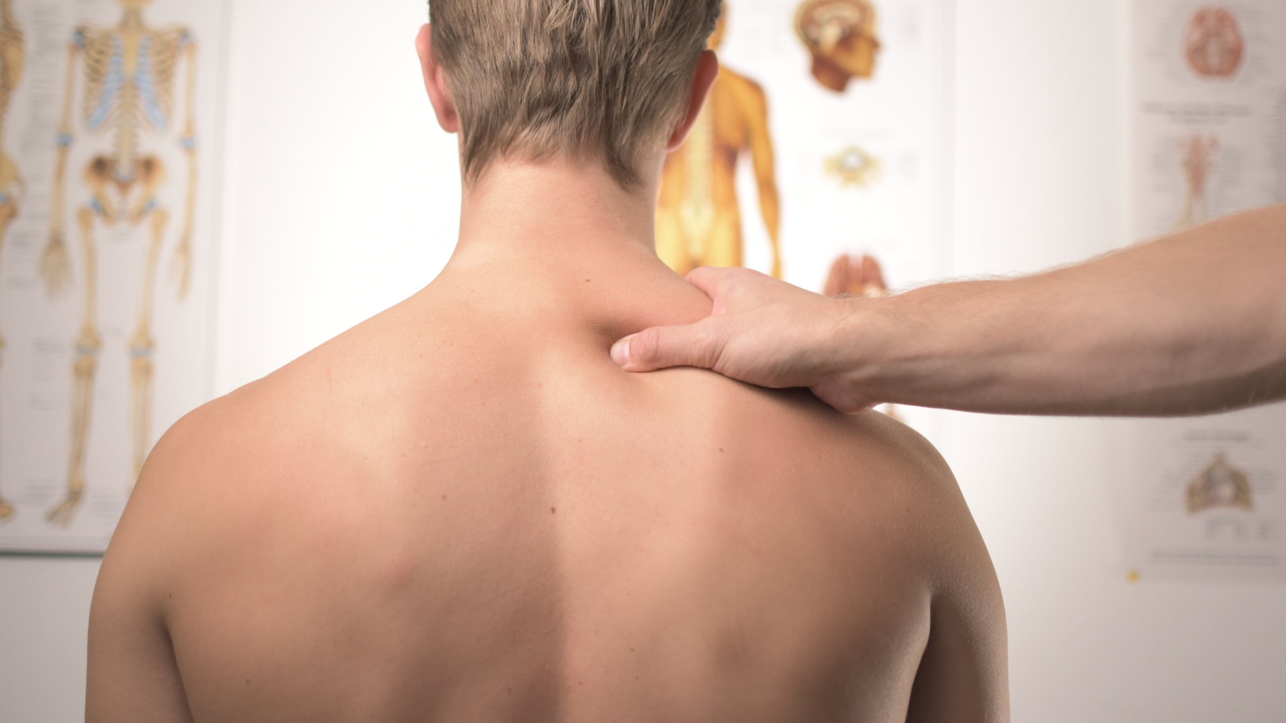
The following is a case study of a patient with complex neck and shoulder pain treated successfully by Jonathan Wray, Principal Physiotherapist at Next Wave in Hilton, WA.
Abstract
Cervical radiculopathy is a condition that affects the cervical nerve roots. The clinical presentation can include neck pain, upper limb pain, sensory changes, paraesthesia, motor deficits or any combination of these. Limited evidence exists as to the best management approach whilst guidelines are based on clinical expertise and patient preference. Surgery is recommended in the presence of persistent severe pain or progressive motor deficits although physiotherapy has been shown to be as effective at longer-term comparison. This case study describes a 43 year old female who presented with a 7 week history of right neck pain, upper limb pain, absent triceps reflex, loss of strength in the C7 myotome and reduced cervical active range of motion. An MRI identified a C6/7 disc extrusion compressing the right C7 nerve root. Management included a combination of manual therapy, exercise and pain neuroscience education. The patient attended for 14 treatments over 12 weeks with substantial improvements measured on the Neck Disability Index, Patient Specific Functional Scale and muscle strength. This case study demonstrates the effectiveness of physiotherapy in managing cervical radiculopathy in a cost efficient and timely manner.
Introduction
Cervical radiculopathy (CR) is a condition resulting from foraminal encroachment of the cervical nerve roots (1). The clinical manifestations of cervical radiculopathy are variable and include neck pain, upper limb pain, sensory changes such as paraesthesia and numbness, motor deficits, diminished reflexes or any combination of these (2). A common cause of foraminal encroachment in CR is cervical spondylosis (1). Degenerative changes at the facet joints affect the posterior aspect of the nerve root and the uncovertebral joints the anterior aspect. Degenerative loss of disc height with associated arthritic joint changes can reduce the diameter of the neural foramen contributing to nerve root impingement (3). Nerve roots C6 and C7 are most commonly affected (2). Herniation of the cervical disc is only responsible in 25% of cases (4). Other causes include trauma but these are relatively uncommon (4). Nerve root compression in isolation does not necessarily lead to radicular pain unless the dorsal root ganglion is involved (1). Some evidence suggests that inflammatory mediators released by the cervical discs during the degenerative process are responsible for symptoms of radicular pain (5).
The age adjusted incidence is approximately 83 per 100,000 persons making it considerably less common than lumbar radiculopathy (1.44 per 100 person) (6)(7). There is limited evidence that documents the natural history of CR (2). A systematic review by Wong et al (8) that evaluated untreated CR concluded most patients report substantial improvements 4 to 6 months after onset and such improvements are maintained over 2-3 years. However, there is a poorer prognosis for those with workers compensation claims (8)(2).
Conservative management for CR includes manual therapy, manipulation, traction, exercise, pharmacology and cervical steroid injection (2). Authors have reported good to excellent results with conservative management (9). However, an estimated 26% of patients with CR are suggested to require surgery (4). Surgery should be considered if pain persists after 6 to 12 weeks of conservative treatment or if there is evidence of progressive neurological deficit (10). Additional features that may warrant early surgical intervention include myelopathy or signs of cervical instability (2).
There are no studies that directly address the question of whether surgery, non-surgical treatment, or a wait and see approach should be used to manage patients with CR. Clinical guidelines have been based on clinical expertise, indirect evidence as well as patient preference (10). National Guidelines for the non-operative management of CR from North America based on low quality evidence or consensus from a systematic review (11) support the use of patient education, non steroidal anti-inflammatory medication, individualized physical activity, motor control exercise, manual therapy and traction. Which has been well described.
This case study describes a CR presentation that reflects the need for a clinician to utilize clinical reasoning guided by the available evidence, whilst incorporating patient preference to achieve optimal clinical outcomes. The patient provided informed consent prior to writing this case study.
Case Study
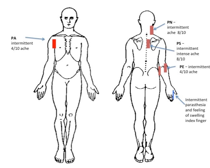
A 43 year old female office worker presented with a 7 week history of right neck pain, upper limb pain, and paraesthesia extending to the level of the right index finger. She reported intermittent pain of up to 8 on a 0-10 numerical rating scale (Figure 1).
Figure 1. Body Chart
(Main complaint: PN, PS, and index finger paraesthesia)
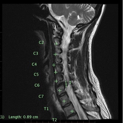
Symptom onset was insidious, and the patient attributed this to poor posture sitting at work. Her symptoms exacerbated 2 weeks after the onset after she fell on steps. An MRI and CT scan organized by the treating General Practitioner (GP) identified a disc herniation at C6/7 (Figure 2). At the time of the initial consultation, the patient was unable to work due to her symptoms and she was worried about the financial implications. She was married with 2 teenage children and prior to injury she enjoyed yoga and walking. The patient was fearful of her diagnosis and long-term implications, and she was quite distressed by other findings on her imaging reports, such as, “multi-level disc degeneration” and “degenerative disc disease”. The patient had attended for other interventions such as chiropractic, massage, acupuncture and Bowen therapy, which had not afforded symptom relief nor provided a long-term strategy for managing her condition. She was using pain medication as directed by her GP, which included paracetamol, Lyrica and Celebrex. An appointment with a neuro-surgeon was pending.
Figure 2. MRI of the cervical spine.
Radiologist report indicated multilevel established cervical disc degenerative changes together with a sizeable broad-based right posterolateral disc extrusion at C6/7 that obscured and likely significantly compressed the exiting intraspinal right C7 nerve root.
Aggravating factors were holding the right upper limb on the steering wheel while driving associated with weakness and production of PN and PS after 5 minutes. Computer work using a mouse reproduced PN, PS and a sensation of swelling in the right index finger after 15 minutes. The patient reported weakness if she maintained her upper limb in these positions and when lifting a washing basket (see Patient Specific Functional Scale, Figure 3). She avoided activities of daily living with her right upper limb. Easing factors were to avoid the aggravating positions, use of anti-inflammatory medication (Celebrex), and greatest relief from lying prone over a gym ball in cervical spine flexion. Her physiotherapy goals were to gain help in resolving her condition without surgery and to get “strong again”. She was concerned that her neck and upper limb pains would prevent her from achieving this and that her current presentation would become chronic.
Physical Examination
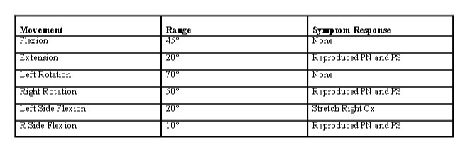
The patient’s usual seated position was slumped with head forward posture. Cervical active range of motion was limited with a consistent pattern of restriction to the right side (see Table 1). All movements were guarded and slow. When low cervical extension was biased, movement was minimal due to guarding and reproduction of PN and PS. Shoulder range of motion was full without pain. Neurological examination demonstrated reduced muscle strength assessed as 4 using a 0-5 scale (American Spine Injury Association) for the C7 myotome, with absent triceps reflex. Palpation over the radial and median nerves demonstrated hyperalgesia in response to palpation at the levels of the proximal upper limb, elbow and forearm, which was not the case for the left side. Neurodynamic testing highlighted mechanosensitivity of the radial and median nerve at 30° of abduction on the right side which was not present when testing the left upper limb. Sensation to light touch and pin prick was normal for the hand and upper limb, although hyperalgesia was noted over the locations of PS and PN. Strength and endurance of elbow extension was assessed using a 2kg weight in supine. The patient was able to achieve 15 repetitions to fatigue on the left side and 5 on the right. This was used as an objective measure to monitor neurological deficit. When the patient moved from supine to sitting she assisted cervical flexion with her hand because she reported her head felt heavy. Craniocervical flexion endurance assessed in supine was 4 seconds. Assessment of shoulder control demonstrated reduced endurance of shoulder flexion at 90° on the right and reduced scapulothoracic control in 4 point kneeling through the right upper limb. This was partly due to reproduction of PS and reporting of weakness by the patient. Spurling’s test was initiated; PN and PS were reproduced with combined cervical right rotation and right side flexion.
Table 1. Showing Cervical Range of Motion Measured with an Inclinometer.
On the first visit the patient completed the Leeds Assessment of Neuropathic Signs and Symptoms questionnaire (S-LANSS) and the short form Orebro (SFO). At the second visit the patient completed the Neck Disability Index (NDI) and Patient Specific Functional Scale (PSFS) as recommended by North American Guidelines (12). On the S-LANSS she scored 6, on the SFO she scored 48/100 and the NDI she scored 18/50, which put her in the moderate disability group (13). The PSFS highlighted difficulty with activities of daily living (see Table 2).
Hypothesis regarding patient’s presentation
Loss of triceps reflex and weakness in the C7 myotome identified during the physical examination combined with the MRI evidence of a C6/7 disc extrusion supported a hypothesis of C7 cervical radiculopathy. Due to the presence of a C6/7 disc extrusion and the fact that the patient gained relief from non-steroidal anti-inflammatory medication, it was hypothesized an inflammatory process was the main cause of her symptoms (5). Neurodynamic and neural tissue provocation testing were indicative of neural tissue mechanosensitivity. The SFO identified an elevated level of concern relating to the long-term prognosis. Interpretation of the NDI and PSFS identified the patients presentation was having a significant impact.
Treatment and Management Plan
Management of this patient began with education on contemporary pain science to help her understand her symptoms and to reduce the fear around them. In a systematic review of Pain Neuroscience Education (PNE) in chronic low back pain, Wood and Hendrick (14) found moderate evidence that PNE added to usual physiotherapy improved disability in the short term. Also discussed were current research findings relating to CR and the favourable natural prognosis to reduce her concern as highlighted on her SFO (15) (4).
Manual therapy was provided initially to reduce symptoms of neural tissue mechanosensitivity, to increase cervical spine active range of motion and to reduce neck pain. This approach has been shown to be affective in individuals with CR (16).
Manual therapy techniques included:
-
Cervical lateral oscillatory glides at C6 to reduce neural tissue mechanosensitivity.
-
Manual cervical traction at C6 to reduce pain.
An exercise program was commenced to retrain motor control of the cervical spine and right upper limb. This consisted of:
-
Cervical rotation in supine with head supported to reduce axial compressive loading of the cervical spine. This exercise was used to address bracing of the cervical musculature and improve active range of motion. The patient was instructed not to move into a provocative range.
-
Deep cranial cervical flexion in supine was deemed appropriate as it had a pain relieving affect and reduced endurance was noted in the physical examination.
-
Cervical extension motor control in 4 point kneeling was given with the patient moving from a fully flexed cervical position i.e. head down, to a neutral cervical position whilst demonstrating optimal cranio-cervical control. This was for postural endurance in a neutral cervical spine position and for weight bearing control through her right upper limb.
-
Supine bilateral shoulder flexion was given for neural mobilization as it was less provocative than abduction and because it related to functional restrictions identified in the physical exam.
-
A supine triceps extension exercise performed with a 2kg dumbbell was given as an objective measure of progression or worsening of neural deficit but also to encourage strength retraining of muscle fibres as neuromuscular activity started to resolve.
Table 2.
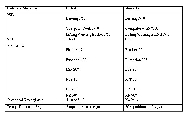
The patient was treated 14 times over 12 weeks during which time her exercise program was progressed. This progression was to target reduced endurance and control of the upper limb and cervical spine in functional positions i.e. using a computer mouse, moving from supine to sitting and lifting up to 5kg. Exercises included:
-
Standing resisted shoulder flexion and extension for right upper limb functional deficits highlighted in physical exam.
-
Cranio-cervical endurance combined with moving supine to sitting.
-
Progression of cervical motor control combining co-ordination of the cervical extensors and cranio-cervical flexors in a neutral spine position whilst adding upper cervical rotation. This was performed in an elbow propped position.
-
Bicep curl and shoulder press combined with a split squat to target lifting related activities.
During this time the patient had a review with a neurosurgeon who made a case for surgical intervention primarily based on her motor deficits. This alarmed the patient, as her preference was a non-surgical approach.
Discussion
Over a 12-week treatment period and 14 sessions of physiotherapy this patient made significant improvements in NDI, PSFS, active range of motion and motor weakness being treated with a combination of manual therapy and therapeutic exercise.
Limited high quality evidence is available on best management strategies for patients with CR (11) (2). This can lead to confusion both for the referring GP and for the patient, which may lead to conflicting advice given by health care providers. Some reports suggest that surgery is most effective if conducted within 12 weeks from onset of symptoms (1). Others suggest a shorter time frame of 8 weeks (7) and that the longer the duration of symptoms the poorer the clinical outcomes are after surgery (20). This may put pressure on primary health care providers and patients to proceed to surgery sooner rather than later due to fear of not achieving optimal outcomes. However apart from progressive neurological deficit, indications for surgery include failure of resolution of symptoms with a conservative approach (1). With regard to early surgical intervention due to motor deficit, Overdevest et al (21) found surgical intervention demonstrated faster improvements in motor weakness in lumbar disc herniations. However this improvement was no longer significant after 26 weeks between surgical and non-surgical groups. Imaging has also shown spontaneous resolution of disc herniations and large disc protrusions in the cervical and lumbar spine with clinical improvements correlating with these changes (22).
Definitive treatment protocols for physiotherapeutic management of patients with CR have not been developed and there are deficits in the literature for physiotherapy treatment approaches (12). In a systematic review of the literature, Boyle et al (23) report a general consensus exits in the literature that manual therapy combined with therapeutic exercise is effective in reducing pain and disability whilst increasing function in patients with CR. In this case key attributes of this patient presentation guided the management approach. E.g. her age and absence of other co-morbidity, her S-LANSS score was low indicating a favourable prognosis and higher likelihood of responding to manual therapy (24), her SFO was 48/100, scores greater than 50 indicate a less favourable prognosis (25), and the patient preference was for a non surgical treatment approach. The use of manual therapy in patients with cervicobrachial pain, particularly passive cervical lateral glides of the associated motion segment been shown to be effective in reducing pain and disability (24) (26). The same technique has been shown to improve range of motion in upper limb neurodynamic testing (18) (26). Traction of the involved cervical motion segment has been shown to decrease pain and perceived disability in patients with CR (18). These techniques were chosen as pain relieving techniques and to reduce neural tissue mechanosensitivity. They were then combined with exercises directed at the patient’s functional limitations highlighted in the physical exam and the PSFS. This management strategy was in line with current opinion and the evidence base. It also supports the opinion that delaying surgery beyond the current 12 week time frame may be appropriate.
Conclusion
This case illustrates that physiotherapy combining manual therapy, exercise and PNE was an effective approach in the management of a patient with CR and should be considered prior to surgery.
Written by Jonathan Wray Principal Physiotherapist Next Wave Therapy
Copyright © Next Wave Therapy (2019)
References
1. Carette S, Fehlings MG. Cervical Radiculopathy. N Engl J Med [Internet]. 2005;353:392–9. Available from: http://www.crossref.org/deleted_DOI.html
2. Iyer S, Kim HJ. Cervical radiculopathy. Curr Rev Musculoskelet Med [Internet]. 2016;9(3):272–80. Available from: http://dx.doi.org/10.1007/s12178-016-9349-4
3. Childress M, Becker B. Nonoperative Management of Cervical Radiculopathy. Am Acad Fam Physicians. 2016;93(9):746–54.
4. Rahdakrishnan K, Litchy WJ, O’Fallon WM, Kurland LT. Epidemiology of cervical radiculopathy: a population-based study from Rochester, Minnesota, 1976–1990. Brain. 1994;117(Pt 2):325–35.
5. Kang J, Georgescu H, McIntyre-Larkin L, Stefanovic-Racic M, Evans C. Herniated Cervical Intervertebral Discs Spontaneously Produce Matrix Metalloproteinases, Nitric Oxide, Interleukin 6 and Prostoglandin E2. Spine (Phila Pa 1976). 1995;20(22):2373–8.
6. Corey DL, Comeau D. Cervical radiculopathy. Med Clin North Am [Internet]. 2014;98(4):791–9. Available from: http://dx.doi.org/10.1016/j.mcna.2014.04.001
7. Alentado VJ, Lubelski D, Steinmetz MP, Benzel EC, Mroz TE. Optimal Duration of Conservative Management Prior to Surgery for Cervical and Lumbar Radiculopathy: A Literature Review. Glob Spine J [Internet]. 2014;4(4):279–86. Available from: http://journals.sagepub.com/doi/10.1055/s-0034-1387807
8. Wong JJ, Côté P, Quesnele JJ, Stern PJ, Mior SA. The course and prognostic factors of symptomatic cervical disc herniation with radiculopathy: A systematic review of the literature. Spine J. 2014;14(8):1781–9.
9. Saal J, Saal J, Yurth E. Nonoperative Management of Herniated Cervical Intervertebral Disc with Radiculopathy. Spine (Phila Pa 1976). 1996;21:1877–83.
10. Fehlings MG, Tetreault LA, Riew KD, Middleton JW, Aarabi B, Arnold PM, et al. A Clinical Practice Guideline for the Management of Patients With Degenerative Cervical Myelopathy: Recommendations for Patients With Mild, Moderate, and Severe Disease and Nonmyelopathic Patients With Evidence of Cord Compression. Glob Spine J. 2017;7(3_supplement):70S–83S.
11. Kjaer P, Kongsted A, Hartvigsen J, Isenberg-Jørgensen A, Schiøttz-Christensen B, Søborg B, et al. National clinical guidelines for non-surgical treatment of patients with recent onset neck pain or cervical radiculopathy. Eur Spine J. 2017;26(9):2242–57.
12. Bono C, Chiselli G, Gilbert T, Kreiner S, Reitman C, Summers J, et al. North American Spine Society Diagnosis and Treatment of Cervical Radiculopathy from Degenerative Disorders. North American Spine society. 2010.
13. Vernon H, Mior S. The Neck Disability Index: A Study of Reliability and Validity. J Manipulative Physiol Ther. 1991;14:409–15.
14. Wood L, Hendrick P, Quraishi N. A systematic review of pain and disability outcomes of pain neuroscience education (PNE) in the management of chronic low back pain. Spine J [Internet]. 2016;16(4):S51. Available from: http://linkinghub.elsevier.com/retrieve/pii/S1529943016001510
15. Health G, Medicine F. Nonoperative Management of Cervical Radiculopathy. 2016;
16. Langevin P, Desmeules F, Lamothe M, Robitaille S, Roy J-S. Comparison of 2 Manual Therapy and Exercise Protocols for Cervical Radiculopathy: A Randomized Clinical Trial Evaluating Short-Term Effects. J Orthop Sport Phys Ther [Internet]. 2015;45(1):4–17. Available from: http://www.jospt.org/doi/10.2519/jospt.2015.5211
17. Murphy DR, Hurwitz EL, Gregory A, Clary R. A Nonsurgical Approach to the Management of Patients With Cervical Radiculopathy: A Prospective Observational Cohort Study. J Manipulative Physiol Ther. 2006;29(4):279–87.
18. Cleland JA, Whitman JM, Fritz JM, Palmer JA. Manual Physical Therapy, Cervical Traction, and Strengthening Exercises in Patients With Cervical Radiculopathy: A Case Series. J Orthop Sport Phys Ther [Internet]. 2005;35(12):802–11. Available from: http://www.jospt.org/doi/10.2519/jospt.2005.35.12.802
19. Epstein N. Iliac crest autograft versus alternative constructs for anterior cervical spine surgery: Pros, cons, and costs. Surg Neurol Int [Internet]. 2012;3(4):143. Available from: http://surgicalneurologyint.com/surgicalint-articles/iliac-crest-autograft-versus-alternative-constructs-for-anterior-cervical-spine-surgery-pros-cons-and-costs/
20. Burneikiene S, Nelson EL, Mason A, Rajpal S, Villavicencio AT. The duration of symptoms and clinical outcomes in patients undergoing anterior cervical discectomy and fusion for degenerative disc disease and radiculopathy. Spine J [Internet]. 2015;15(3):427–32. Available from: http://dx.doi.org/10.1016/j.spinee.2014.09.017
21. Overdevest GM, Vleggeert-Lankamp CLAM, Jacobs WCH, Brand R, Koes BW, Peul WC. Recovery of motor deficit accompanying sciatica – Subgroup analysis of a randomized controlled trial. Spine J [Internet]. 2014;14(9):1817–24. Available from: http://dx.doi.org/10.1016/j.spinee.2013.07.456
22. Casey E. Natural History of Radiculopathy. Phys Med Rehabil Clin N Am [Internet]. 2011;22(1):1–5. Available from: http://dx.doi.org/10.1016/j.pmr.2010.10.001
23. Boyles R, Toy P, Mellon J, Hayes M, Hammer B. Effectiveness of manual physical therapy in the treatment of cervical radiculopathy: a systematic review. J Man Manip Ther [Internet]. 2011;19(3):135–42. Available from: http://www.tandfonline.com/doi/full/10.1179/2042618611Y.0000000011
24. Nee RJ, Vicenzino B, Jull GA, Cleland JA, Coppieters MW. Baseline Characteristics of Patients With Nerve-Related Neck and Arm Pain Predict the Likely Response to Neural Tissue Management. J Orthop Sport Phys Ther [Internet]. 2013;43(6):379–91. Available from: http://www.jospt.org/doi/10.2519/jospt.2013.4490
25. Linton SJ, Nicholas M, MacDonald S. Development of a short form of the Örebro musculoskeletal pain screening questionnaire. Spine (Phila Pa 1976). 2011;36(22):1891–5.
26. Hall TM, Elvey RL. Nerve trunk pain: physical diagnosis and treatment. Man Ther. 1999;4(2):63–73.